Direct Ophthalmoscopes
In direct ophthalmoscopy, an ophthalmoscope is used to examine the fundus from a close distance. The examiner can view the central parts of the eye – such as the optic nerve papilla, vascular sources and the macula lutea (yellow spot). Since the magnification used in direct ophthalmoscopy is higher than that used in indirect ophthalmoscopy, this procedure allows the examiner to see more detail, but they can only view a small section of the fundus two-dimensionally.
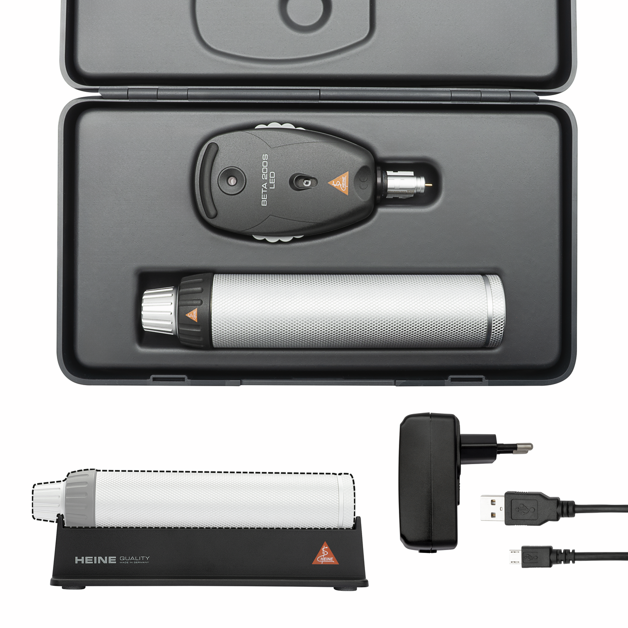
These examinations are particularly successful with the instruments included in our HEINE BETA 200 range and featuring aspherical optics, preferably with LEDHQ. But the HEINE K180 or the two mini 3000 ophthalmoscopes (with LEDHQ or XHL illumination) are ideal too.
These examinations are particularly successful with the instruments included in our HEINE BETA 200 range and featuring aspherical optics. But the HEINE K180 or the mini 3000 ophthalmoscope are ideal too. All the instruments feature LEDHQ illumination for precise and uniform illumination of the examination area, plus true-to-life colour rendering when looking through the ophthalmoscope.
The BETA X Ophthalmoscope with the AOS+ optical system offers additional examination functions through its swivel-in polarisation filter and focusCONTROL.
Direct ophthalmoscopes are often used in the early detection of ocular disorders in children (Brückner Test).
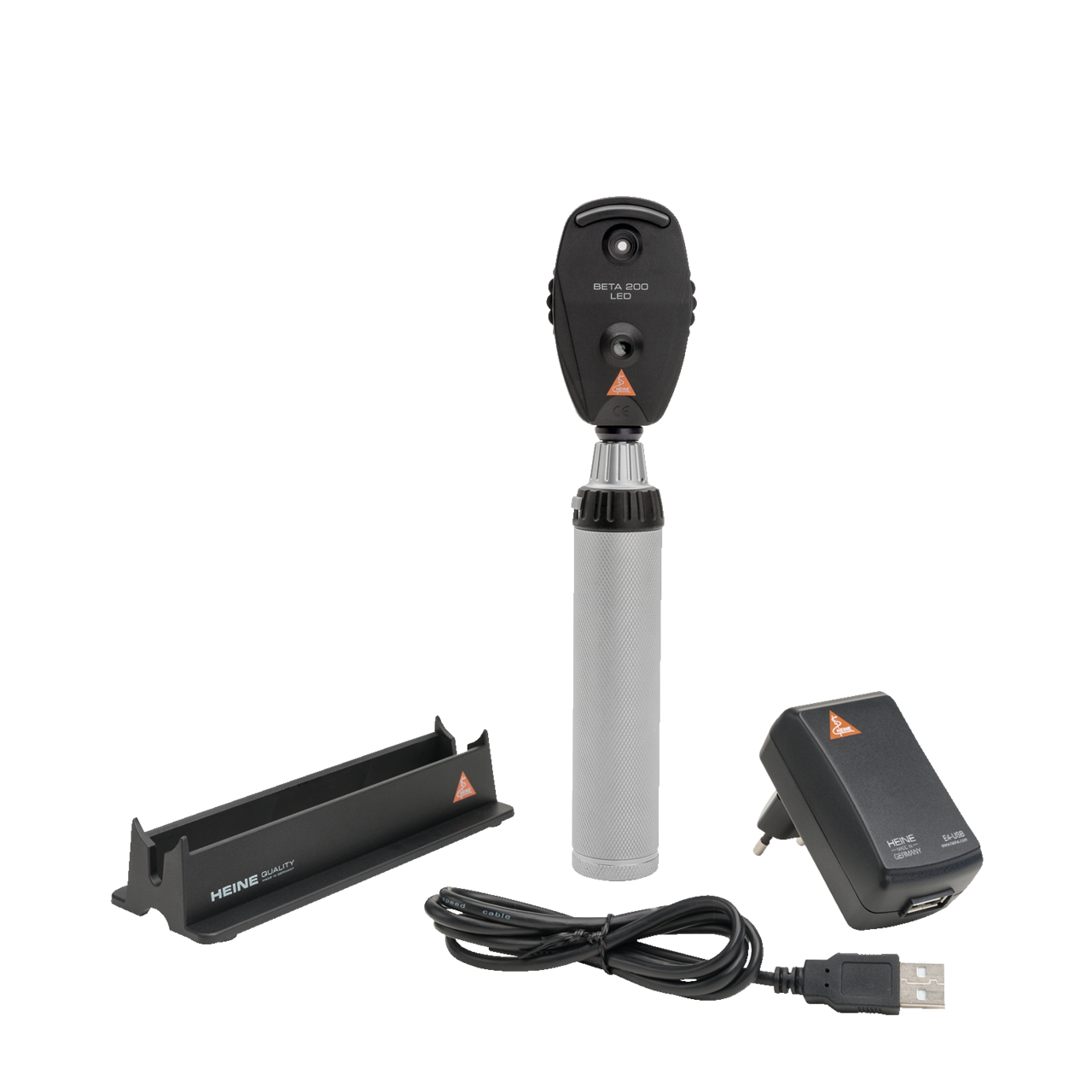
Ophthalmoscope with outstanding durability
Ophthalmoscope with outstanding durability – perfect for small pupils
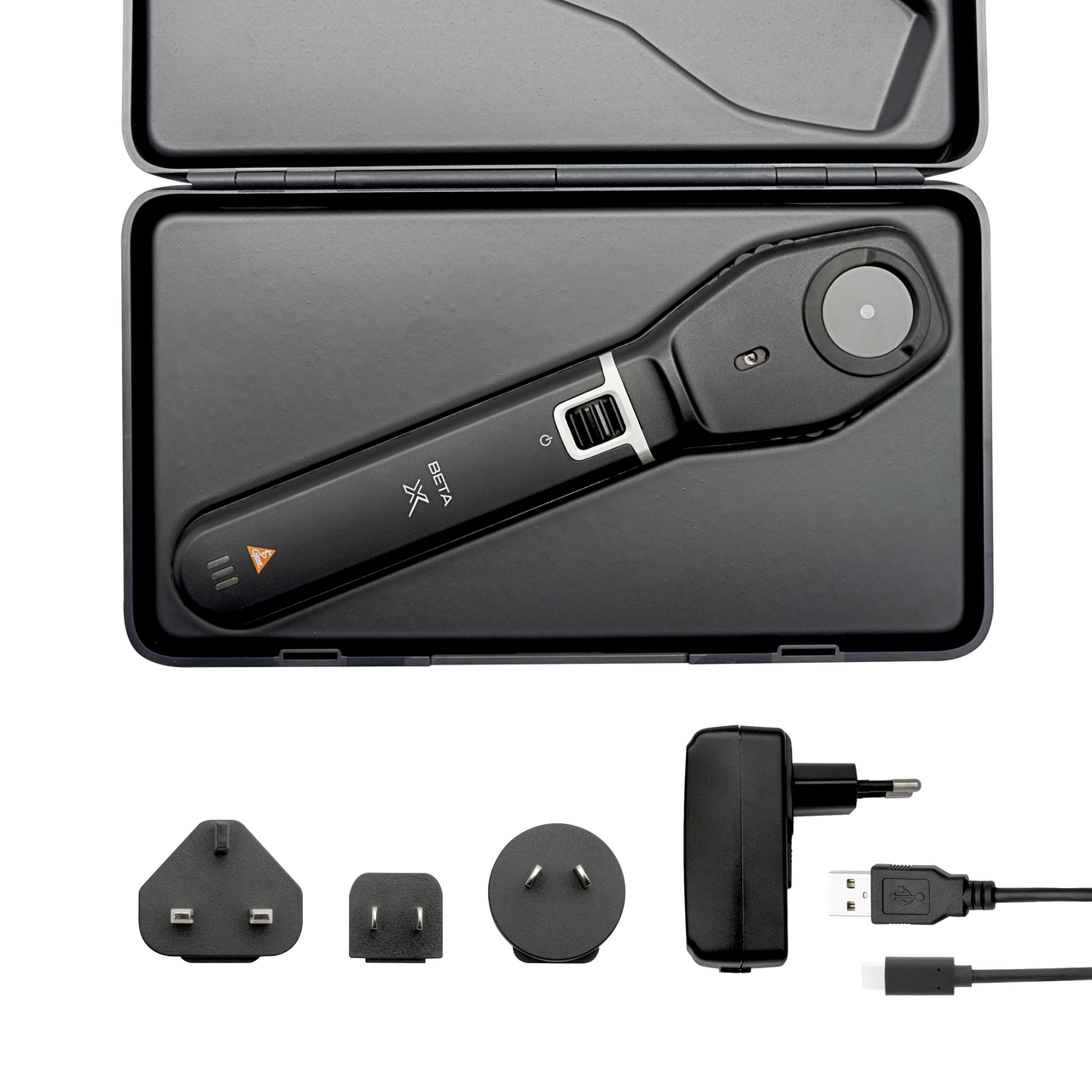
Our most advanced direct ophthalmoscope
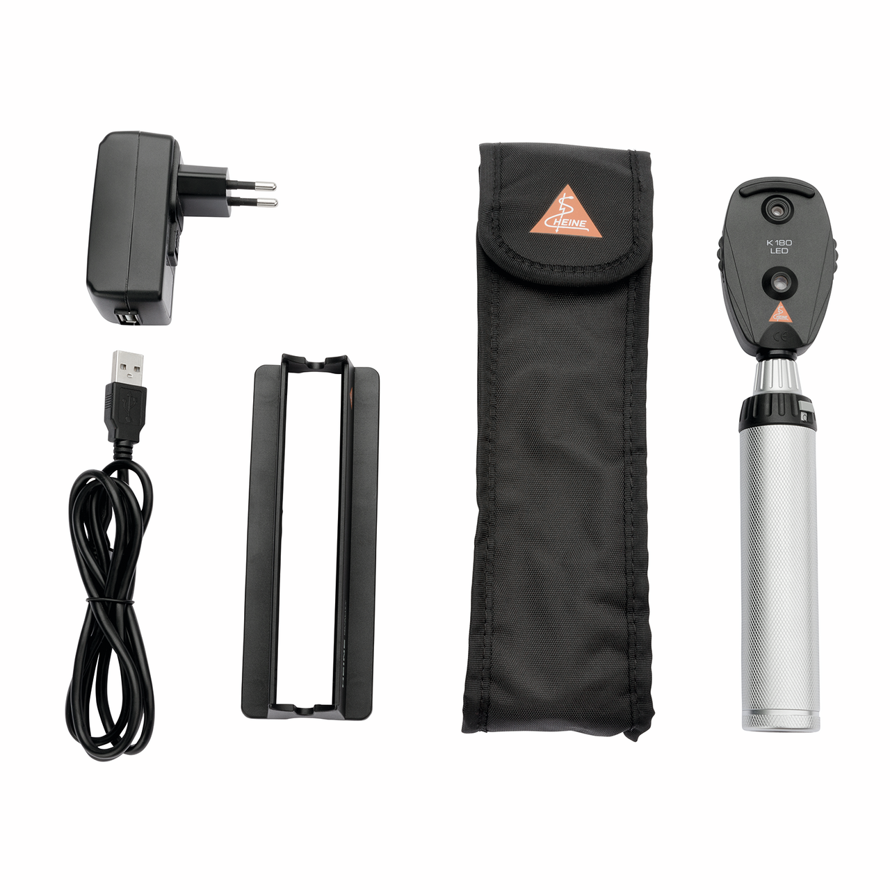
Standard instrument for a smaller budget
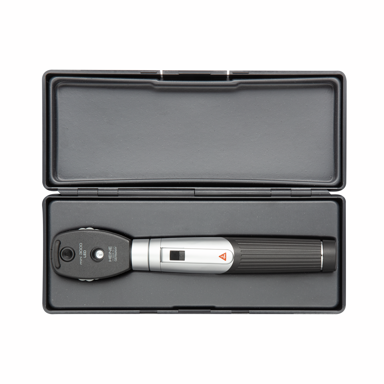
Maximum quality with minimum dimensions