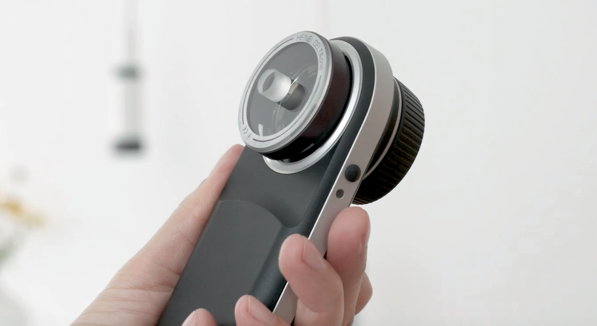
A small contact plate makes diagnosing easier
Dermatoscopy of lesions between fingers and toes
The dermatoscope is considered to be the most important instrument in the dermatologist’s auxiliary equipment toolkit. It has become a vital part of the process of reliable assessments of unpigmented and pigmented lesions, such as naevus cell naevi and melanomas in the context of skin cancer screening. But the dermatoscope is also an important instrument when it comes to detecting other lesions such as plantar warts, hypertrophic sebaceous glands or itch mite infestations. However, reaching prominent places with a dermatoscope is not always straight forward. Often, the clinician cannot get close enough with the dermatoscope in contact mode to sufficiently examine some areas of the skin like for example in interdigital areas, in the ear, in the corners of the eyes or the nasal wings.
In situations such as this it is advisable to use a smaller contact plate like the one available for the HEINE DELTAone or DELTA 30 dermatoscopes. The narrow optical attachment sits perfectly in concave-shaped body parts, therefore enabling dermatoscopy there too.
It is impractical in particular to examine lesions between the toes using a normal dermatoscope in contact mode. Pigmented lesions such as naevus cell naevi (which require clarification) are frequently found here too. A dermatoscope with a small contact plate is ideal for easily distinguishing Verrucae vulgares (plantar warts) which also develop on the sides of the toes from foreign bodies or calluses (corns).
Dermatoscopic facial examinations
A smaller dermatoscopic plate is the perfect partner for examining exposed parts of the face – the nose wings, inside and behind the auricle, as well as in the visible external ear canal – Dermatologists often observe white skin cancer (such as basal cell carcinomas) or precancerous white skin cancer (including actinic keratoses) on sun-damaged skin in these areas.
Purely cosmetic lesions such as hypertrophic sebaceous glands, xanthelasma or milia often occur on the forehead, in the middle of the face, in the inner corners of the eyes, on the eyelids or in the nose area. A small, suitable dermatoscope attachment is helpful when dealing with these localised areas which can be difficult to examine with a standard dermatoscope, particularly in light of the fact that these lesions can clinically resemble a basal cell carcinoma, a white skin cancer that is frequently found at the nasal wings or corners of the eyes.
Scabies: on the trail of the itch mite
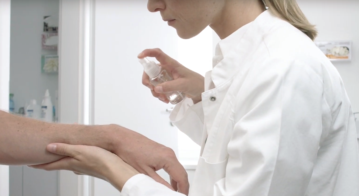
Itchy red papules, blisters and scratch marks are all indications of an itch mite infection. Itch mites – tiny spider-like creatures that dig corridors 0.5 to 2 millimetres wide under the skin – like to nest in the spaces between the fingers or in the navel. In this situation, a more acccurate diagnosis can be made with a small contact plate used in direct contact with the skin and with immersion fluid. This then allows a dermatologist to reliably distinguish an itch mite infestation from other disease patterns such as eczema in the context of neurodermatitis.
Immersion fluid provides a helping hand in diagnosis
It is advisable to use an immersion fluid in non-polarised examination mode. The immersion fluid ensures that the dermatoscope’s light is directed to the skin. Therefore deeper structures of the skin become more visible. The immersion fluid balances out differences between the skin and the contact plate, creating a more even image without any annoying air pockets which can make diagnosis difficult.
Some dermatoscope manufacturers offer smaller contact plates as optional accessories. HEINE Optotechnik provides contact plates as an accessory in its range for the DELTA 30 and DELTAone precision optical dermatoscopes. The small disc measures a mere 8 millimetres in diameter, it can be used quickly and easily by simply clicking onto the head of the dermatoscope.