Diagnostics with the indirect ophthalmoscope
Applications in practice
Many ophthalmologists work with the slit lamp. So the greater resolution undoubtedly produces better images of central detailed findings in the macula area or on the optic nerve, for example. But, when it comes to having an overview of and screening the periphery, the head-worn indirect ophthalmoscope makes the examination process faster and gives you a greater overview.
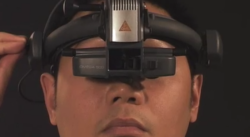
Fast, mobile and at a distance – performing a retinal exam with the indirect ophthalmoscope
Examining the retina with the binocular indirect ophthalmoscope (BIO) is a more patient and practitioner-friendly method compared to an examination involving the slit lamp – especially during the contiuing coronavirus crisis when we’re supposed to avoid contact where possible, carry out exams quickly and maintain social distancing. If we follow these steps, we can assume that transmission of infectious diseases is less likely.
The benefits of the head-worn indirect ophthalmoscope both in a hospital setting and for large practices are obvious: the BIO can be taken wherever needed, giving the ophthalmologists much flexibility. For example, in-depth retinal examinations can be conducted on wheelchair-using or recumbent patients who cannot sit at the slit lamp as well as small children .
As an ophthalmologist working in private practice, the everyday demands are different from those you’d encounter in a hospital setting. Here, the most important factors are time, patient comfort and the quality of the exam.
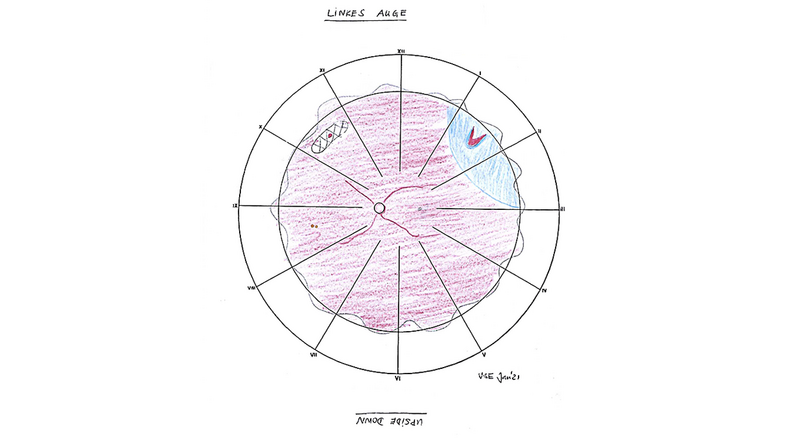
An exam performed with the binocular indirect ophthalmoscope can tick all these boxes – and provide comprehensive diagnostic information to boot. It gives an examiner a very quick overview of the entire fundus – into the far periphery – in 3D (with scleral indentation if necessary). In addition, the lower magnification of the BIO (binocular indirect ophthalmoscope) usually improves visualisation if there are media opacities in the eye, such as advanced cataracts.